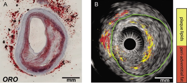
Figure 4: Ex Vivo Lipid Differentiation Result of the Atherosclerotic Left Anterior Ascending Coronary Artery of an 80-year-old Man.

(A) Histology: Oil Red O (ORO) staining of the intravascular photoacoustic/intravascular ultrasound image in cross-section. The lipids are shown in red. (B) Lipid differentiation map overlaid on the co-registered intravascular ultrasound image of the coronary artery. The lipids in the plaques are yellow and lipids in the peri-adventitial tissue are red. The green contour indicates the external elastic lamina. Adapted from Wu et al., 2015.[49]
