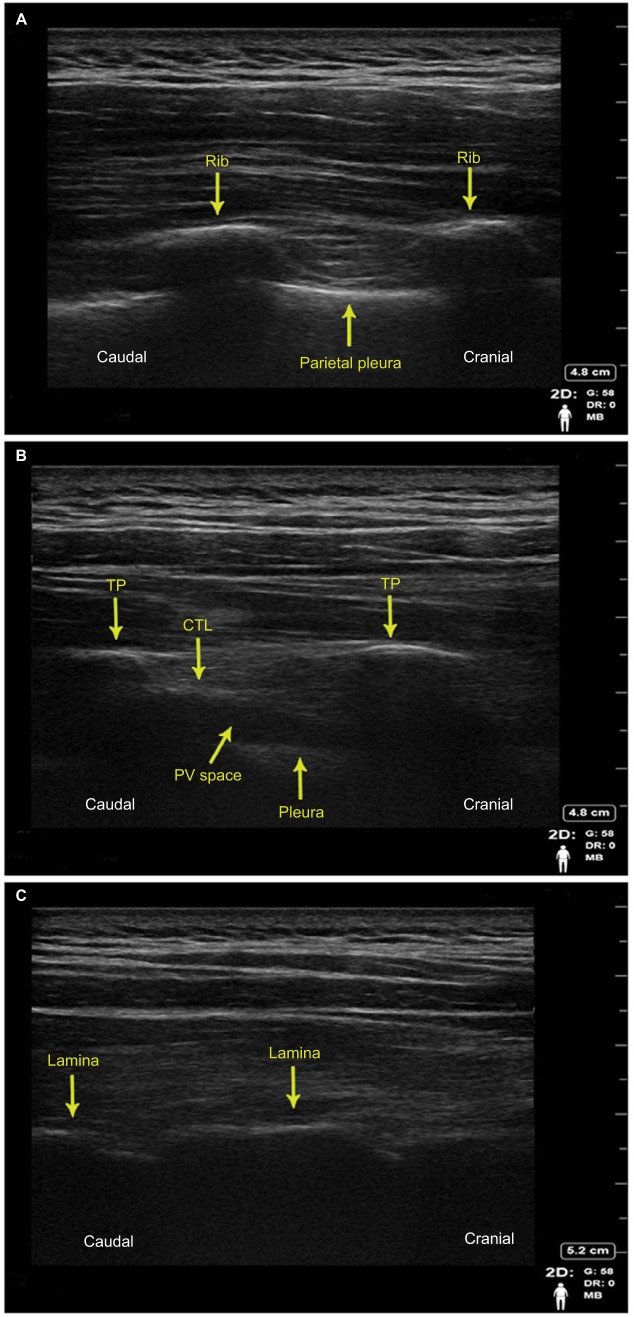
Figure 2.
Human sonoanatomy images obtained by paramedian sagittal plane ultrasonography in a healthy volunteer, sitting position, X-Porte ultrasound machine, HFL50XP 15-6 MHz linear transducer (SonoSite).
Notes: (A) Proximal intercostal view; (B) “Classical” PV; and (C) Retrolaminar view.
Abbreviations: TP, transverse process; CTL, costotransverse ligament; PV, paravertebral.

