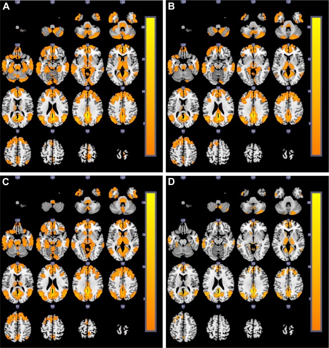
Figure 1.
The cross-sectional MRI images of brain activation regions (yellow areas) related to DMN (the left posterior cingulate cortex as the seed) among four groups.
Notes: (A) Control subjects; (B) mild COPD group; (C) moderate COPD group; (D) severe COPD group.
Abbreviations: DMN, default mode network; MRI, magnetic resonance imaging.

