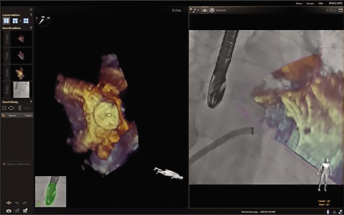
Figure 5: EchoNavigator® Release II During Left Atrial Appendage Occlusion Procedures.

Integration of echo information into the X-ray image after left atrial appendage (LAA) closure. The new release of the EchoNavigator allows for exact delineation of the LAA morphology after device implantation. The opacification of the echo overlay can be reduced in order to more or less delineate the radiolucent device position.
