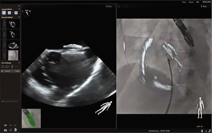
Figure 6: EchoNavigator® During Closure of Interatrial Septum Defects.

The translation of echo information into the fluoroscopic image allows for safer device implantation. In this example one can reproduce the correct position of the occluder device in the echocardiographic image. The overlay image enables precise judgement of the relation between the occluder device and the delivery catheter after release of the system in real time.
