
Figure 11.
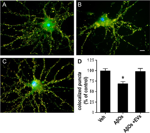
MSC-derived EVs protect hippocampal neurons from synapse damage induced by AβOs. Representative double immunolabeling images for synaptophysin (red) and PSD-95 (green) in cultured hippocampal neurons exposed to vehicle (A), 500 nm AβOs for 24 h (B), or 500 nm AβOs plus EVs (2.4 × 10⁸ particles) (C). EVs were incubated with neuronal cultures for 22 h at 37 °C following initial exposure to AβOs (500 nm) for 2 h. Nuclei are shown in blue (DAPI) and co-localization of pre- and postsynaptic markers is shown in yellow. Images were acquired on a Zeiss Axiovert 200M microscope with a ×63/1.25 numerical aperture oil objective. Scale bar, 10 μm. D, synapse density determined as the number of co-localized synaptophysin/PSD-95 punctae, normalized to vehicle (Veh)-exposed cultures. Bars, means ± S.E. (error bars) (n = 2 independent cultures, with triplicate coverslips per experimental condition). *, p < 0.05; one-way ANOVA followed by Dunnett's post hoc test.