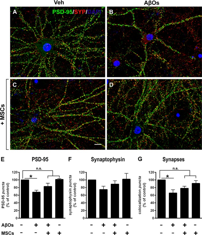
Figure 3.
Levels of pre- and postsynaptic proteins in hippocampal neurons exposed to AβOs. A–D, representative photomicrographs showing double immunolabeling for presynaptic marker synaptophysin (SYP, red) and postsynaptic marker PSD-95 (green) in hippocampal neurons exposed to vehicle or AβOs (500 nm) for 24 h; nuclei are stained by DAPI (blue). SYP/PSD-95 co-localized puncta (a measure of synapse density) appear in yellow. Scale bar, 10 μm. Images were acquired in a Nikon Eclipse TE300 epifluorescence microscope with a ×63 objective. E–G, quantification of synaptic proteins and synapse density. Data are represented as means ± S.E. (error bars) (n = 4 independent cultures, with triplicate coverslips in each experimental condition); *, p < 0.05; two-way ANOVA followed by Dunnett's post hoc test; n.s., not significant.

