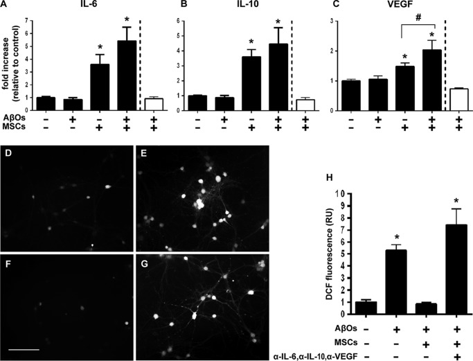
Figure 7.
Secretion of IL-6, IL-10, and VEGF to the culture medium is increased in neuronal/MSC cocultures and mediates neuroprotection against AβO-induced oxidative stress. A–C, levels of IL-6 (A), IL-10 (B), and VEGF (C) in the culture medium are increased when hippocampal neurons are in coculture with MSCs, either in the absence or presence of AβOs. No changes in cytokine levels are detected when MSCs alone are exposed to AβOs (white bars, normalized by control MSCs in the absence of AβOs). D–H, the addition of antibodies against IL-6 (0.5 μg/ml), IL-10 (1 μg/ml), and VEGF (1 μg/ml) to the culture medium blocks the protection by MSCs against AβO-induced neuronal oxidative stress. D and E, representative DCF fluorescence images from hippocampal neuronal cultures exposed to vehicle or 500 nm AβOs, respectively. F and G, representative DCF fluorescence images from hippocampal neurons cocultured with MSCs and incubated with 500 nm AβOs in the absence or presence of anti-cytokine antibodies, respectively. Images were acquired on a Nikon Eclipse TE300 epifluorescence microscope with a ×20 objective. Scale bar, 100 μm. H, integrated DCF fluorescence intensities in different experimental conditions, normalized by the control group (vehicle-exposed hippocampal neurons in the absence of MSCs). Bars, means ± S.E. (error bars) (n = 2 independent cultures, with triplicate wells per experimental condition). *, p < 0.05; #, p < 0.05; one-way ANOVA followed by Tukey's post hoc test.

