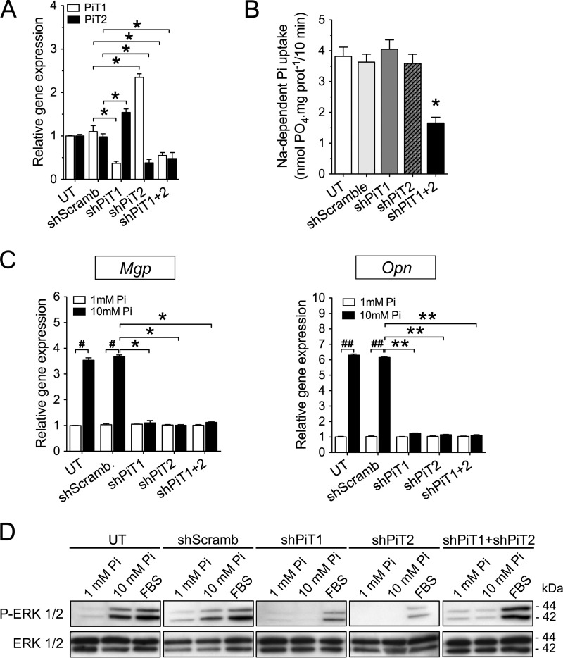
Figure 1.
Pi-dependent Mgp and Opn gene regulation and ERK1/2 signaling require both PiT1 and PiT2 in MC3T3-E1 cells. A, RT-qPCR analysis of PiT1 (white bars) and PiT2 (black bars) mRNA levels in untransfected (UT) or stably transfected MC3T3-E1 cells, as indicated. Data are means ± S.E. (*, versus shScramb, p < 0.05, n = 3). B, sodium-dependent Pi uptake was measured in untransfected or stably transfected MC3T3-E1 cells, as indicated. Data are means ± S.E. (n = 3). C, RT-qPCR analysis of Mgp and Opn mRNA levels in untransfected or stably transfected MC3T3-E1 cells, as indicated. Cells were incubated in low-serum (0.5%) medium for 24 h and stimulated with 1 mm (white bars) or 10 mm (black bars) extracellular Pi concentration for 24 h. Data are means ± S.E. (error bars) (#, p < 0.05; ##, p < 0.01 versus 1 mm Pi control; and *, p < 0.05; **, p < 0.01 versus shScramb; n = 3) D, Western blot analysis of ERK1/2 phosphorylation (P-ERK 1/2) in untransfected or stably transfected MC3T3-E1 cells, as indicated. Cells were incubated in low-serum (0.5%) medium for 24 h and stimulated for another 24 h with 1 mm or 10 mm extracellular Pi concentration or with 10% FBS used as a positive control for ERK1/2 phosphorylation. Total ERK1/2 proteins were used as a loading control.

