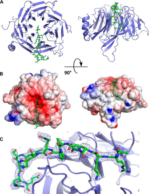
Figure 2.

Crystal structure of RBBP4 in complex with BCL11A(2–16) peptide. RBBP4 is shown as a cartoon in blue, and BCL11A(2–16) is shown as sticks in green. A, BCL11A(2–16) is bound to the top face of the RBBP4 β-propeller and then proceeds to wrap down the side. B, electrostatic surface potential representation of RBBP4–BCL11A(2–16). The surface is colored according to the calculated electrostatic potential from −8 kT/e (red) to 8 kT/e (blue). C, 2Fo − Fc electron density map of the BCL11A(2–16) peptide contoured at 1σ is shown as blue mesh.
