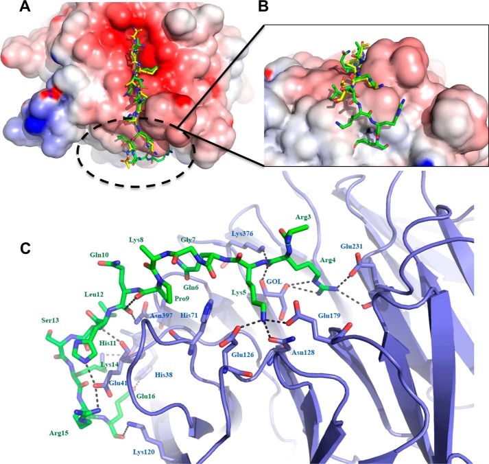
Figure 3.
Comparison of histone H3 and BCL11A peptides bound to RBBP4. A, overlay of histone H3 peptide (shown in yellow) (20) (PDB code 2YBA chains B and C) and the BCL11A peptide (shown in green) bound to the top face of the RBBP4 β-propeller. B, view of the novel interactions of the BCL11A peptide to the side of RBBP4. C, detailed look at the BCL11A- binding site. Hydrogen bonds are depicted as black dashed lines. Carbon atoms are shown in green for the peptide and slate for the protein; nitrogen atoms are blue, and oxygen atoms are red.

