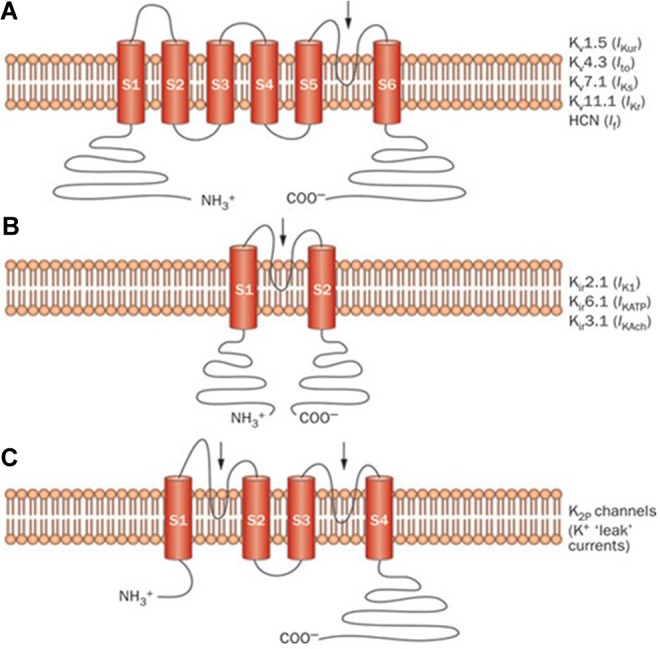
Figure 2.

Structure of different cardiac potassium channel species: Schematic representation of selected potassium channel α-subunits. A, The 6-transmembrane 1-pore-region voltage-dependent K+ channel (Kv) α-subunits mediating IKur, Ito, IKs, IKr, and If. B, The 2-transmembrane 1-pore-region inward rectifying K+ channel (Kir) α-subunits mediating IK1, IKATP, and IKAch. C, The 4-transmembrane 2-pore-region K+ channel (K2P) mediating “leak” K+ currents. The arrows indicate the location of the pore-forming region(s). HCN indicates hyperpolarization-activated cyclic nucleotide-gated channel; If, inward rectifier mixed Na+ and K+ “funny” current; IK1, inward rectifier K+ current; IKACH, acetylcholine-activated inward rectifier K+ current; IKATP, ATP-sensitive K+ current; IKr, rapid component of the delayed rectifier K+ current; IKs, slow component of the delayed rectifier K+ current; IKur, ultrarapid component of the delayed rectifier K+ current; Ito, transient outward K+ current. Reprinted with permission from Giudicessi and Ackerman. Macmillan Publishers Ltd, copyright 2012.3
