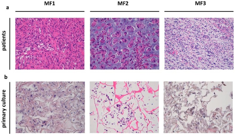
Figure 1.
(a) H&E staining of the surgical specimen showing high-grade myxofibrosarcoma (light blue stroma) infiltrating fibrotic tissue. 20× magnification. (b) H&E staining of MFS primary culture (light blue spots) seeded into tridimensional scaffold. 20× magnification.
H&E, hematoxylin and eosin; MFS, myxofibrosarcoma.

