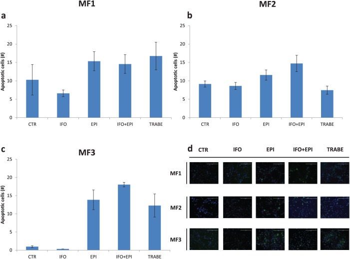
Figure 4.
Number of TUNEL-positive MFS cells in (a) MF1 primary culture, (b) MF2 primary culture and (c) MF3 primary culture. (d) TUNEL staining of MFS primary cultures (10× magnification) showing apoptotic cells (green spots) and cell nuclei (blue spots). Scale bar 400 µm.
MFS, myxofibrosarcoma; TUNEL, terminal deoxynucleotidyl transferase nick end labeling.

