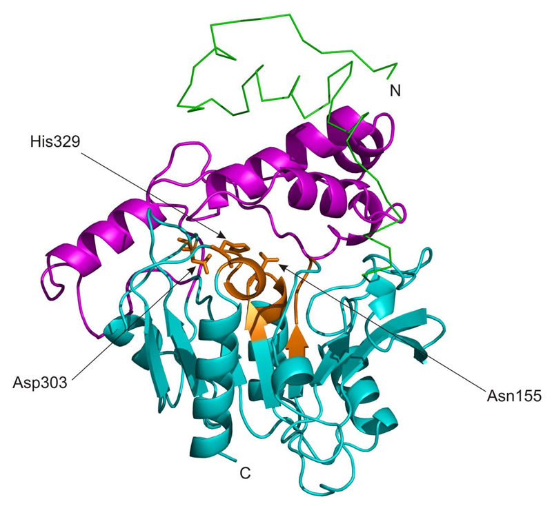
Figure 3.
3D model of murine CGI-58. The αβ hydrolase core structure and the mostly helical cap are shown as cartoon colored in cyan and magenta, respectively. Conserved regions harboring the G-X-S/N-X-G motif, HX4D motif, and the proposed catalytic Asp303 are highlighted in orange. A conserved region around amino acid 86, which is predicted to participate in forming the oxyanion-hole is also colored in orange. The N-terminal extension is displayed as a green line.

