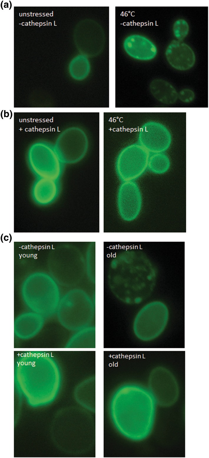
Figure 9.

α‐Synuclein and cathepsin L. fluorescence microscopy of the strain BY4741 pUG35‐α‐synuclein pESC‐HIS. In unstressed cells α‐synuclein‐GFP is closely associated with the plasma membrane. A heat shock (10 min; 46°C) leads to a detachment of α‐synuclein from the plasma membrane and to an accumulation in cytosolic foci (a). Fluorescence microscopy of the strain BY4741 pUG35‐α‐synuclein pESC‐HIS cathepsin L. similar to (a) α‐synuclein‐GFP is localized at the plasma membrane but forms no visible cytosolic aggregates (b). Fluorescence microscopy of the strains BY4741 pUG35‐α‐synuclein pESC‐HIS and BY4741 pUG35‐α‐synuclein pESC‐HIS cathepsin L after elutriation. With and without the expression of cathepsin L α‐synuclein‐GFP is localized at the plama membrane in young cells (fraction II). In old cells (fraction V) α‐synuclein‐GFP detaches from the plasma membrane and forms aggregates. No such aggregates can be seen after expression of cathepsin L and the protein is still visible in the plasma membrane [Colour figure can be viewed at wileyonlinelibrary.com]
