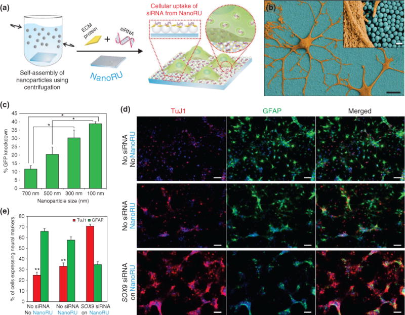
FIGURE 11.

Nanotopography-mediated platform to deliver nucleic acids for stem cell differentiation. (a) Silica nanoparticles (SNPs) are assembled on a film and coated with extracellular matrix (ECM) proteins and nucleic acids (siRNA) to develop the nanotopography-mediated reverse uptake (NanoRU) platform. Neural stem cells (NSCs) cultured on this platform uptake the siRNA, which induces their differentiation into neurons. (b) Scanning electron microscopy image of neurons (brown) with extended axons cultured on NanoRU wherein the SNPs (blue) are visible. (c) Depending on the size of the SNPs on the surface, the uptake rate of siRNA is affected, which is reflected by the difference in green fluorescent protein (GFP) knockdown. Nanoparticle with 100 nm diameter showed the highest efficiency. (d) Fluorescence images showing the differentiation of NSCs into neurons using the NanoRU platform, and (e) the expression of the specific markers, Tuj1 (neuronal) and GFAP (glial cells), was quantified to reveal that NanoRU is an effective platform to induce stem cell differentiation. (Reprinted with permission from Ref 76. Copyright 2013 Nature Publishing Group)
