
FIGURE 3.
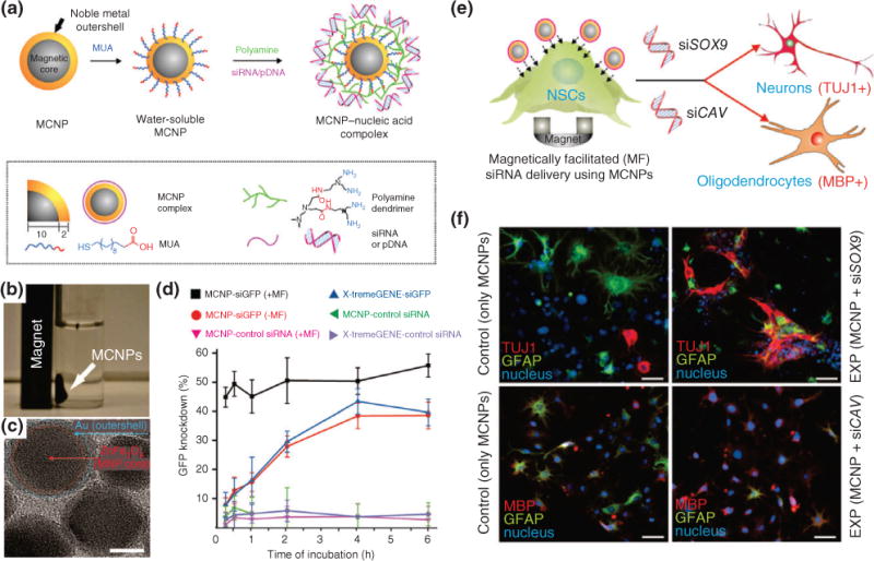
Magnetic core–shell nanoparticles (MCNPs) for stem cell differentiation and imaging. (a) Schematic of MCNPs functionalized with mercaptoundecanoic acid (MUA) followed by electrostatic conjugation of polyamide and nucleic acids for regulating gene expression in stem cells. (b) A representative image showing that MCNPs with a composition of ZnFe2O4 are attracted to a magnet. (c) TEM image of MCNPs (scale bar = 10 nm). (d) MCNPs were incubated in green fluorescent protein (GFP)-labeled rat neural stem cells (rNSCs) and exposed to magnetofection (MF). The resulting GFP knockdown was quantified and is directly correlated to the gene-regulating efficiency of the MCNPs. The greater the GFP knockdown, the greater its effect. (e) Schematic of rNSCs undergoing MF with MCNPs coated with nucleic acids targeting specific stem cell differentiation. (f) Immunofluorescence images showing the differentiation into neurospecific lineages with particular markers, TUJ1 (neuronal), GFAP (glial cells), and MBP (oligodendrocytes), based on the type of nucleic acid delivered. (Reprinted with permission from Ref 38. Copyright 2013 John Wiley and Sons)