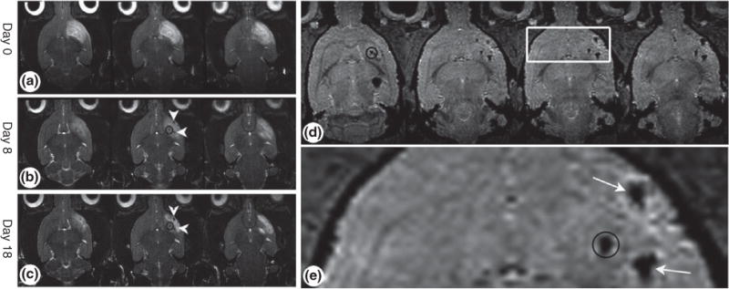
FIGURE 4.

Tracking transplanted stem cells using magnetic resonance imaging (MRI) in vivo. Mesenchymal stem cells (MSCs) were loaded with magnetic nanoparticles (MNPs) and transplanted into rat brains. The loaded MSCs migrate toward the cortical lesion. (a–c) Time course of weighted MRI of rats that were induced with cortical damage. (d) Axial three-dimensional images showing accumulation of MSCs in the cortex and striatum. (e) Enlargement of the white box in (d). Throughout the time period, high-resolution MRI revealed that cells migrated along the distant route toward the lesion. The black circles represent the location of the induced lesion. White arrows point to MNP-loaded MSCs. (Reprinted with permission from Ref 43. Copyright 2008 John Wiley and Sons)
