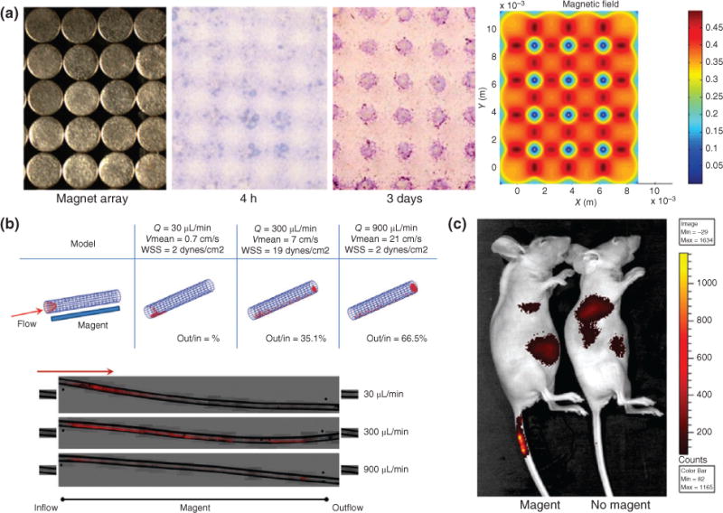
FIGURE 5.

Guiding in vivo localization of magnetic nanoparticles (MNPs) using external magnets. (a) To demonstrate that MNPs are precisely controlled by the location of a magnetic field, a magnet array with spherical patterns was placed underneath a solution of MNPs, and resulted in MNPs localizing to locations of highest magnetic strength. (b) To simulate MNPs flowing in the bloodstream, a MNP solution passed through the tube with a magnet underneath and the localization of the MNP (red) is dictated by the flow rate of the solution. (c) MNPs were intravenously injected into the distal portion of the mouse tail vein while a magnet was placed at the injection site. High signal in the tail vein of the mice with the magnet confirms that localization of MNPs can be externally controlled by a magnet. (Reprinted with permission from Ref 46. Copyright 2013 John Wiley and Sons)
