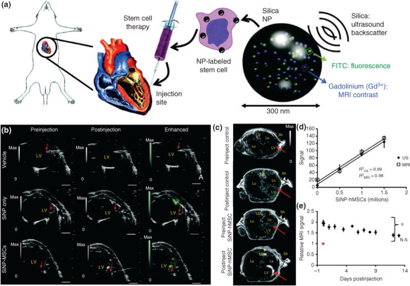
FIGURE 6.

Silica nanoparticles (SNPs) for tracking stem cells in vivo using ultrasound. (a) Schematic of SNPs embedded with FITC for fluorescence imaging and gadolinium for enhancing MRI contrast delivered to mesenchymal stem cells (MSCs) and injected into rat heart tissue. (b) Ultrasound images of human MSCs (hMSCs) after intracardiac implantation in mice. The red arrow represents the bevel of the needle catheter. (c) MRI contract images show enhancement of SNP accumulation. (d) Quantification of the MRI and ultrasound (US) signal as a function of number of injected SNP-loaded hMSCs. (e) Animals injected (on day 0, red dot) with hMSCs were monitored sequentially for 12 days postinjection. (Reprinted with permission from Ref 56. Copyright 2013 The American Association for the Advancement of Science)
