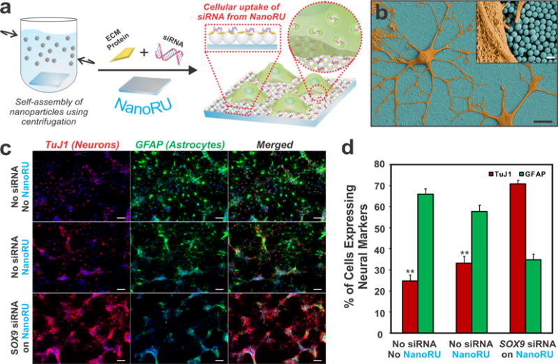
Figure 5.

Nanotopographical features can mediate significant uptake of siRNA by rNSCs. (a) NanoRU facilitates cellular uptake of siRNA into rNSCs. (b) SEM image of rNSCs (orange) on NanoRU (blue). Scale bars: 10 μm and (inset) 500 nm. (c) Images stained for the neuronal marker TuJ1 (red) and the astrocytic marker GFAP (green) showing the extent of differentiation of rNSCs grown on bare glass with no NanoRU or siSOX9 coating (top), NanoRU without the siSOX9 coating (middle), and NanoRU with the siSOX9 coating (bottom). Scale bars: 50 μm. (d) Plot showing quantification of the percentage of cells expressing specific neural markers on various substrates (**, p < 0.001). Adapted with permission from ref 35. Copyright 2013 Nature Publishing Group.
