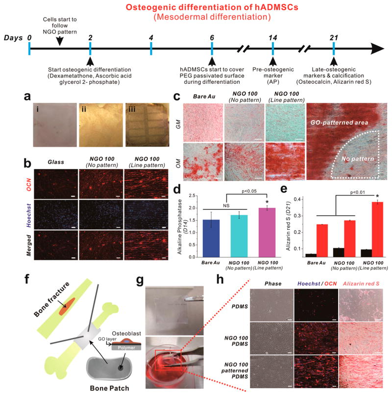
Figure 4.
Osteogenic differentiation of hADMSCs using NGO line-patterned substrate. (a) Picture of (i) glass, (ii) NGO-coated gold substrate, and (iii) NGO line-patterned substrate used for the osteogenesis of hADMSCs for 3 weeks, showing ultimate stability of NGO line pattern generated on the substrate. (b) Fluorescence images of hADMSCs differentiated into osteoblasts stained for osteocalcin (red, top row), nucleus (blue, middle row) and merged (last row), showing elongated and well-spread morphology of hADMSCs that directly followed the geometry of NGO line pattern, which are different from the same cells on glass or NGO-coated substrates. NGO line pattern is clearly visible in Hoechst-stained images in the middle row. Scale bar = 50 μm. (c) Differentiated osteoblasts stained with Alizarin red S at day 21 to confirm the level of calcification (red color) from the cells on bare Au, NGO-coated substrate and the substrate with NGO line pattern. GM and OM mean growth medium and osteogenic medium, respectively. “No pattern” means the area which is not covered by NGO line pattern due to the incomplete transfer of NGO on the surface. Scale bar = 50 μm. (d) Alkaline phosphatase (AP) assay to confirm the expression of preosteogenic marker which is dependent on the type of the substrates. (e) Quantitative analysis of calcium expression of cells treated with GM (black) and OM (red) obtained by extracting Alizarin red S. Results are medians of absorbance signals (460 and 560 nm for AP and Alizarin red S, respectively) obtained from three independent experiments n = 3; *p < 0.05 or p < 0.01, Student’s t-test. “NS” indicating that the two groups were not significantly different at p = 0.05). (f) Schematic illustration showing possible application of proposed “Bone Patch” composed of supporting polymer (PDMS, Young’s modulus: 6.2 MPa), NGO-patterned layer and the osteoblasts derived from hADMSCs from the patient suffering bone fracture. (g) Picture of highly flexible bone patch having NGO pattern and the differentiated osteoblasts. (h) Cells stained for osteocalcin (OCN, red), nucleus (blue) and calcium (Alizarin red S, red) to confirm the successful differentiation into osteoblasts. Scale bar = 50 μm.

