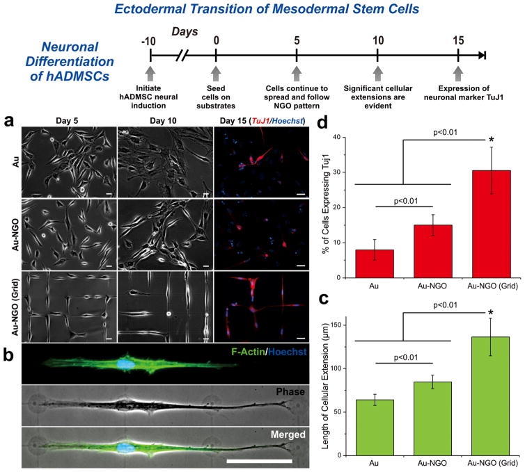
Figure 5.
Neuronal differentiation of hADMSCs using NGO grid-patterned substrate. (a) Images of neural induced hADMSCs grown on PLL-coated Au [Au], NGO-coated Au [Au-NGO], and NGO grid-patterned substrates [Au-NGO (Grid)]. All substrates were coated with laminin to facilitate cell attachment. Cellular growth and morphology were monitored over 15 days, followed by staining for the neuronal marker TuJ1 (red) and nucleus (blue). Scale bars = 20 μm. (b) Phase contrast and fluorescence images of cells stained for F-actin (green) and nucleus (blue) after 15 days of cultivation show extensive cellular extension on NGO-grid patterns. Scale bar = 50 μm. (c) Quantitative comparison of the length of cellular extension on various substrates (n = 3; *p < 0.01, Student’s unpaired t test). (d) Quantitative comparison of the percentage of cell expressing the neuronal marker TuJ1 on various substrates (n = 3; *p < 0.01, Student’s unpaired t test).

