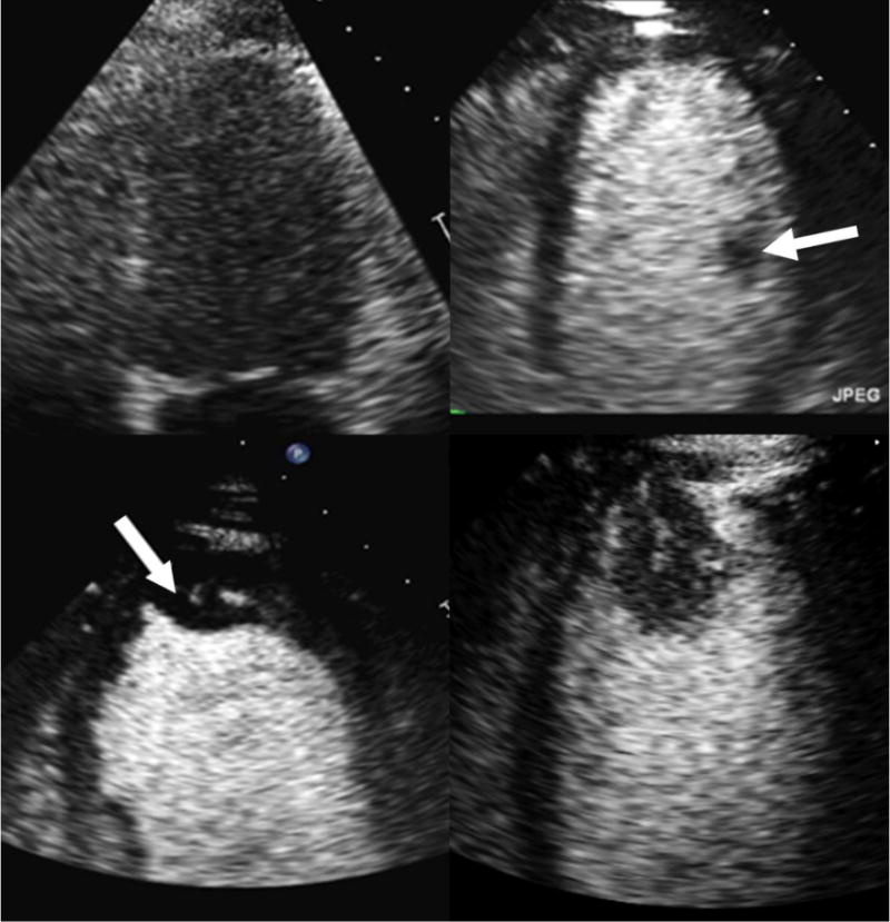
Figure 3.

Endocardial borders are not well visualized without contrast (top left). Contrast microbubbles opacify the left ventricle improving delineation of endocardial borders (top right). A papillary muscle (white arrow), a normal intracardiac structure, is outlined by the contrast. Contrast enables clear visualization of an apical thrombus (arrow; bottom left) and apical tumor (bottom right). The presence of a vascular channel and enhancement with contrast help to differentiate apical tumor from apical thrombus.
