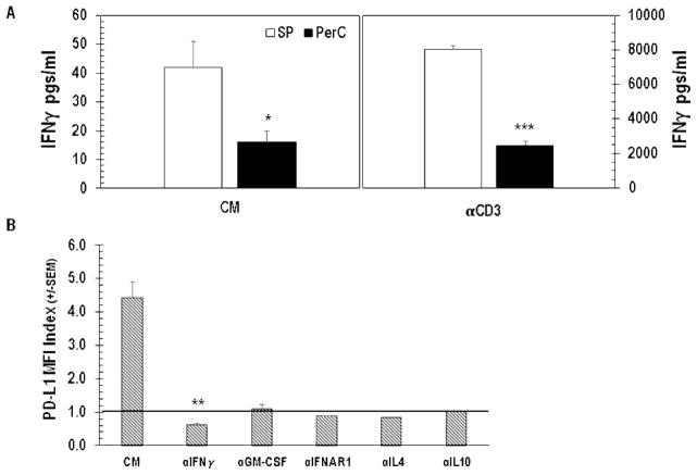
Figure 4.
IFNγ drives PD-L1 expression. Panel A: Unstimulated (CM) and stimulated (αCD3) SP or PerC cell cultures were screened for IFNγ production by ELISA. Results depict averages from 6 experiments. Panel B: PerC cells cultured without (CM) or with neutralizing mAbs to the cytokines listed were analyzed for PD-L1 expression after 48 hrs. Data from 3–5 experiments.

