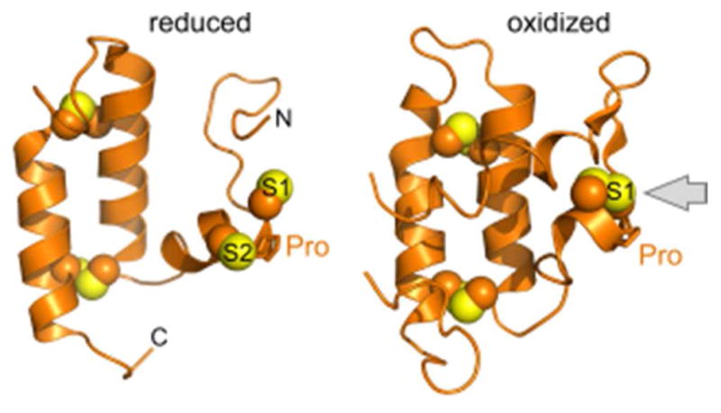
Figure 12.

Structures of the Mia40 oxidoreductase. Sulfurs in the CPC motif are labeled S1 and S2. Gray arrow in the oxidized structure illustrates solvent accessibility of the redox-active disulfide. PDB codes are 2K3J (reduced; H. sapiens Mia40 solution NMR structure) and 2ZXT (oxidized; S. cerevisiae Mia40 crystallized as a fusion with maltose binding protein (not shown)).
