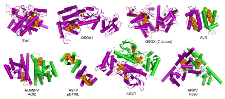
Figure 17.
Gallery of sulfhydryl oxidase flavoenzyme structures. Protein subunits are purple and green, FAD is orange, disulfide bond sulfurs are yellow. Ero1, Saccharomyces cerevisiae Ero1; QSOX1, Rattus norvegicus Quiescin Sulfhydryl Oxidase 1; QSOX (T. brucei), Trypanosoma brucei Quiescin Sulfhydryl Oxidase; ALR, Homo sapiens Augmenter of Liver Regeneration; AcMNPV, Autographa californica multicapsid nucleopolyhedrovirus; ASFV, African swine fever virus; AfGliT, Aspergillus fumigatus Gliotoxin Sulfhydryl Oxidase; APMV, Acanthamoeba polyphaga mimivirus. With the exception of APMV R596, dimer structures are viewed down the two-fold axis. PDB codes are Ero1, 1RP4; QSOX1, 4P2L; QSOX (T. brucei), 3QCP; ALR, 1OQC; AcMNV Ac92, 3QZY; ASFV pB119L, 3GWL; AfGliT, 4NTC; APMV R596, 3GWN.

