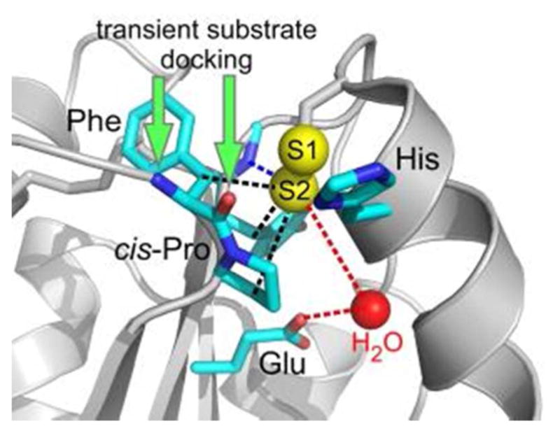
Figure 8.

Representative active site of a PDI family protein. Amino acids commonly observed in PDIfamily active sites are shown in stick representation, with red indicating oxygen atoms and blue nitrogen. The Phe side chain is replaced by Tyr in some cases. Potential interactions of the largely buried S2 sulfur of the CXXC motif are indicated by dashed lines (black—hydrophobic; blue—hydrogen bonding; red— proton transfer). Green arrows indicate sites for hydrogen bonding interactions by the incoming substrate backbone. Red sphere is water. PDB code is 3ED3 (yeast Mpd1p).
