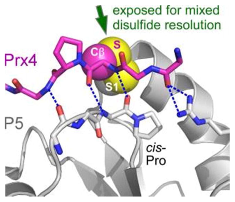
Figure 9.

Model for substrate interaction with a PDI protein. The P5 protein (gene name PDIA6) is in gray, and the backbone (including a proline side chain) of a peptide from peroxiredoxin Prx4 is in magenta. The cysteine side chain atoms of Prx4 are labeled Cβ and S. Blue dashed lines are hydrogen bonds. Red in stick representations indicates oxygen atoms, blue nitrogens. The S1 sulfur of P5 is labeled. PDB code is 3W8J.
