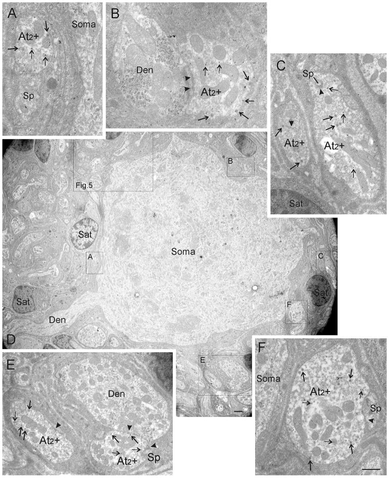
FIGURE 4.
Ultrastructure of CG cells receiving GABA-immunogold-stained At2 inputs. D shows a low magnification overview of a typical CG neuron whose perisomatic neuropil is exclusively supplied by GABA-positive terminals as indicated by the presence of immunogold particles overlaying them (thin arrows). These were classified as At2 terminals (At2 +, boxes A–F) by using the formerly defined criteria, (e.g. the high number of dense-core vesicles [arrows, A–F]) and less asymmetric synaptic densities (arrowheads). Detailed views of immunogold-labeled At2 terminals labeled as boxes in D are shown in A, B, C, E, and F. A higher magnification electron micrograph of the big box is shown in Figure 5A. Abbreviations: At2 + = axon terminal type 2 positively immunogold-stained for GABA, Den = dendrite, Sat = satellite cell, Sp = spine. Scale bar = 0.5 μm in A–C, E–F; 2.0 μm in D

