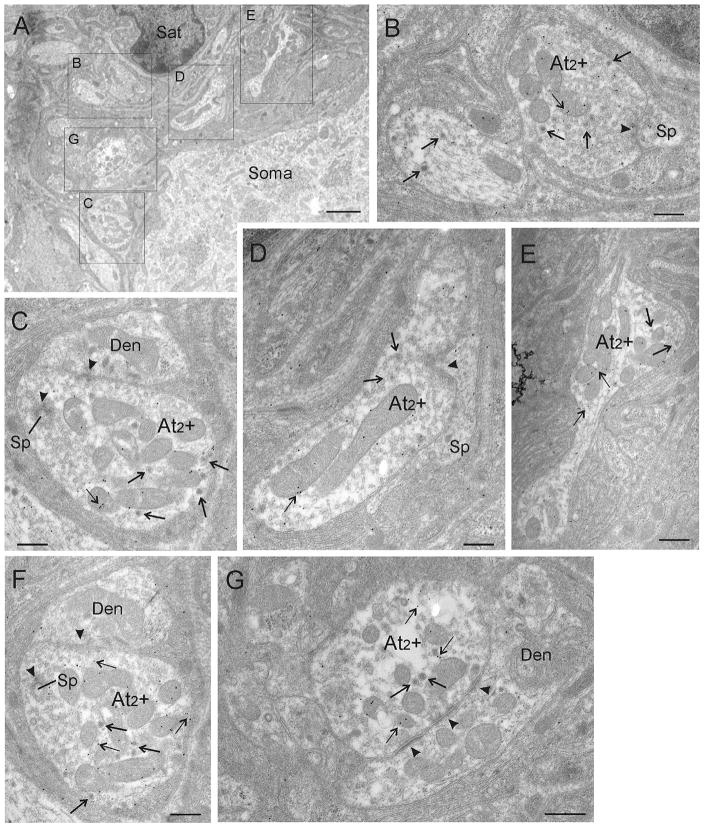
FIGURE 5.
A. Ultrastructural details of GABA-positive terminals. Higher magnification photomicrograph of the big box in Figure 4D showing another set of GABA-positive At2 + terminals supplying the same CG neuron. B–G. High magnifications are shown of the terminals indicated by labeled boxes in A. Several immunogold-particles (small arrows) overlie each At2 terminal, indicating that At2 terminals are GABA-positive (B–G). Images in C and F show the same At2 terminal in adjacent serial sections. Abbreviations: At2 + = axon terminal type 2 positively immunogold-stained for GABA, Den = dendrite, Sat = satellite cell, Sp = spine. Scale bar = 2.0 μm in A; 0.5 μm in B–G

