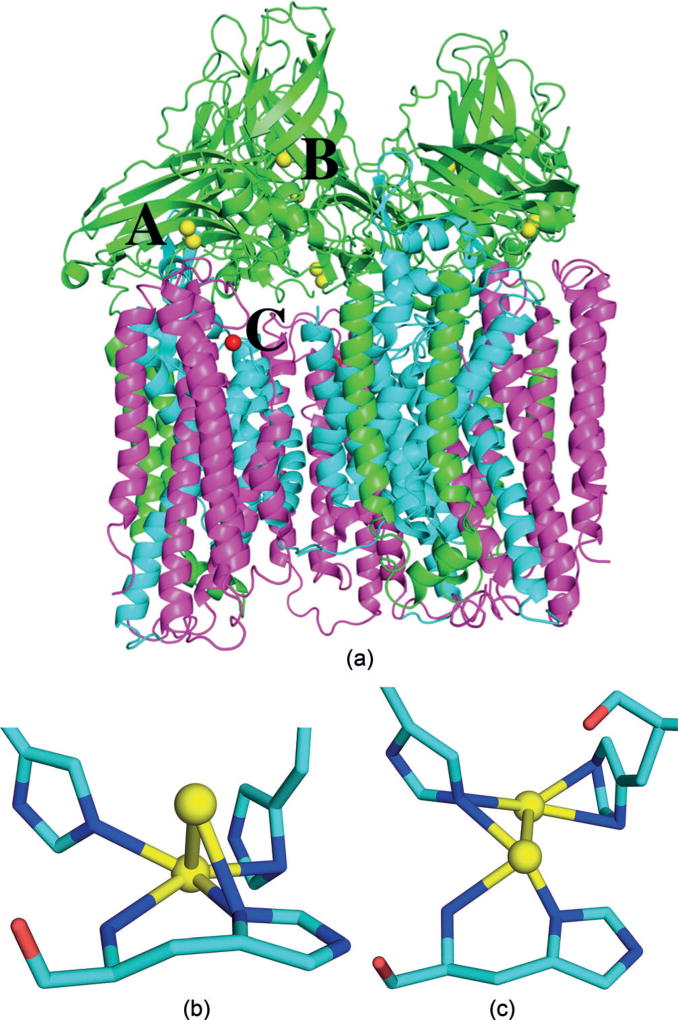
Figure 1.
a) Trimeric structure of the pMMO (3RGB)[9] with metal sites A–C indicated. Cu ions in yellow, Zn ions in red; PmoA, PmoB, and PmoC subunits in magenta, green, and cyan, respectively. Geometry of Cu site A in protomers 1 (b) and 2 (c), involving three histidine residues and the amino terminal group. Cu ions are shown as yellow balls whereas C, N, and O atoms are shown as cyan, blue, and red sticks, respectively.

