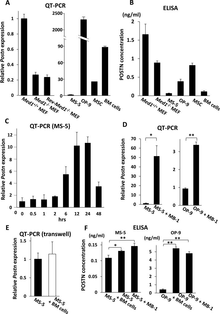
Fig. 3. POSTN expression in various mesenchymal cells and BM cells.
(A) Quantitative PCR. Postn mRNA was reduced in Med1−/− MEFs compared with Med1+/+ MEFs, and was not restored in Rev-Med1−/− MEFs (left panel). Postn was variably expressed in BM stromal cells and BM cells (right panel). The values are plotted as the fold increase versus the value in Med1+/+ MEFs (left panel) or MS-5 cells (right panel).
(B) ELISA of culture media. POSTN was secreted by various mesenchymal cells and BM cells.
(C–E) Quantitative PCR. MS-5 cell Postn mRNA was prominently induced when cocultured with BM cells (C). MS-5 cell or OP-9 cell Postn mRNA was induced when cocultured with MB-1 cells for 24 h (D). When cocultured for 24 h in transwell to inhibit direct contact between BM cells and MS-5 cells, Postn mRNA was not induced (E).
(F) ELISA of culture media. mPOSTN secretion was induced by physical interactions of MS-5 or OP-9 cells with BM cells or MB-1 cells.
N = 3 (A–F).

