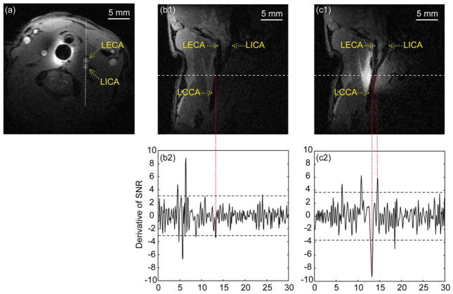
FIG. 5.
(a) Axial slice to locate carotid bifurcation when the passive resonator is inserted inside the esophagus. The acquisition parameters are TR/TE=163.3/2.5 ms, 35° flip angle, 3×3cm2 FOV, 0.8-mm slice thickness, 256×256 matrix, and number of acquisitions (NA)=2. The dashed line passing through the internal and external carotid arteries defines the orientation of longitudinal images to be acquired subsequently. (b1) Longitudinal slice acquired with flow saturation in the absence of pumping power. The acquisition parameters are TR/TE=348.5/4.3 ms, 32° flip angle, 3×3cm2 FOV, 0.4-mm slice thickness, 300×300 matrix, and NA=4. (c1) Longitudinal slice acquired in the presence of pumping power, with other acquisition parameters the same as (b1). The bandwidths for all three images are 25 kHz. (b2) and (c2) are derivative plots of 1D intensity profiles normalized to the same noise floor when plotted along the dashed lines in (b1) and (c1) and the horizontal dashed lines correspond to twice the SD evaluated over each derivative plot. LECA, left external carotid artery; LICA, left internal carotid artery; LCCA, left common carotid artery.

