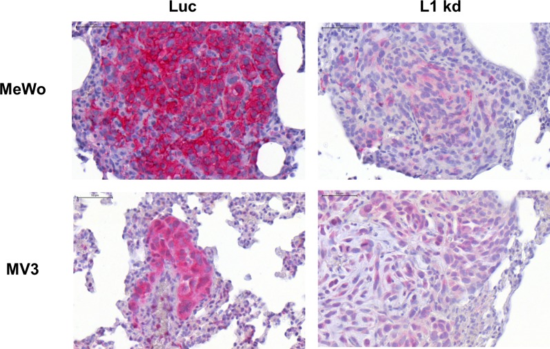
Fig 3. L1CAM expression in lung metastases from human melanoma xenograft as detected by immunohistochemical analysis.
Immunohistochemical staining for L1CAM expression (red) in lung metastases of melanoma cells MeWo (upper panels) and MV3 (lower panels) with unchanged L1CAM expression (Luc, right panels) and L1CAM knockdown (L1 kd, left panels). All scale bars: 50 μm. Please note that staining was amplified for MV3.

