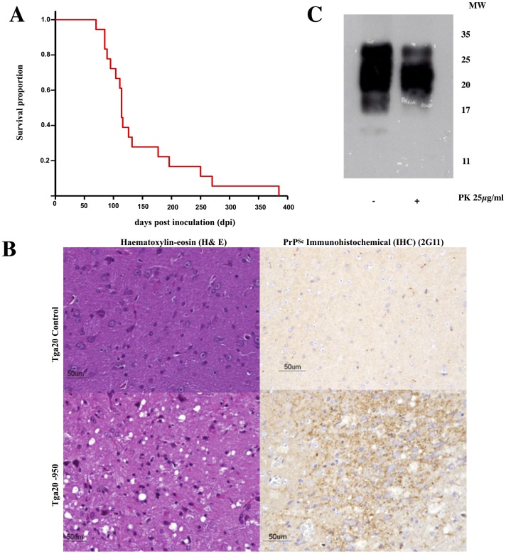
Fig 2. Characterization of the infectivity of recPrPSc.
A) Kaplan-Meier survival plots of Tga20 mice inoculated with recMoPrPSc. B) Histopathological and immunohistochemical analysis of brains fromTga20 mice inoculated with recMoPrPSc and uninoculated tga20 controls: left: haematoxylin-eosin (H&E) staining of the medulla oblongata, notice the spongiform lesion in the inoculated mice (bottom); right: PrPSc IHC staining (antibody 2G11, epitope: 151–159) of the medulla oblongata showing fine granular PrPSc deposits in the inoculated mice (bottom). C) WB showing the presence of PK-resistant PrP in the brains of Tga20 mice inoculated with recMoPrPSc.

