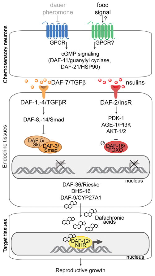
Figure 2. Role of insulin/IGF-1 and TGFβ pathways in controlling reproductive growth versus dauer development.
The pathways are depicted under favorable conditions in which the concentration of dauer pheromone is low (grayed out) and the concentration of an uncharacterized food signal is high. Under these conditions, the food signal is thought to promote GPCR signaling in chemosensory neurons, leading to DAF-11/guanylyl cyclase activity and increased levels of cGMP, resulting in secretion of TGFβ and insulin ligands. DAF-7/TGFβ binds to DAF-1,-4/TGFβR, which regulates Smad/co-Smad transcriptional complexes. Insulins bind to DAF-2/InsR, leading to phosphorylation by AKT-1/2 of DAF-16/FOXO, which is retained in the cytosol. TGFβ and insulin signaling promotes the biosynthesis of the dafachronic acids, which bind to DAF-12/NHR in target tissues that functions transcriptionally to promote reproductive growth. Under unfavorable conditions in which dauer pheromone is high and food signal is low, chemosensory neurons do not secrete insulin and TGFβ ligands. DAF-16/FOXO becomes dephosphorylated, translocates into the nucleus, and acts as a transcription factor, while DAF-3/Co-Smad and DAF-5/Sno/Ski are no longer inhibited by DAF-8/-14 and thus can act as a transcription factor. Consequently, the biosynthesis of the dafachonic acids is inhibited, unliganded DAF-12 binds to its inhibitor DIN-1 and promotes a transcriptional program that triggers dauer formation.

