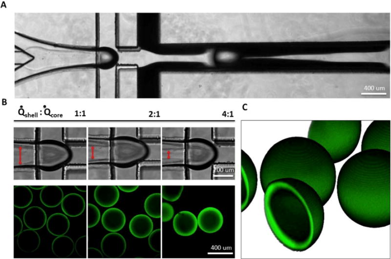
Figure 2. Microcapsule fabrication.

A) Coaxial flow-focusing generates hollow core-shell capsules. B) Shell thickness (visualized with FITC-PEG-SH) was varied by modulating core/shell flow rate ratio. C) 3D reconstruction of z-stacked confocal microcapsule images. Capsule diameter is ~400 μm, shell thickness is ~10 μm.
