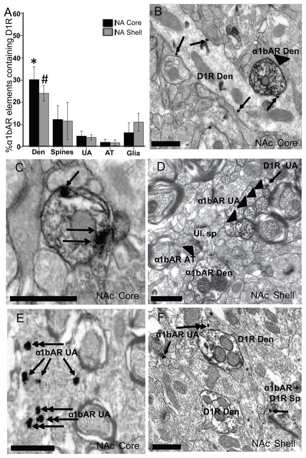
Figure 2.
α1bAR and D1R display a low degree of colocalization in the core and shell of the NAc. (A) The mean percent (± SEM) of α1bAR immunoperoxidase-containing elements that also contained immunogold labeling representing the D1R in both the core and shell of the NAc. *p<0.01 indicates in the NAc core, a significantly greater percentage of double-labeled dendrites vs. unmyelinated axons, axon terminals and glial elements. #p<0.05 indicates in the NAc shell, significantly more double-labeled dendrites than unmyelinated axons and axon terminals. (B) Representative electron micrograph from the NAc core with an immunoperoxidase-labeled dendrite representing α1bAR (arrowhead) and a D1R labeled dendrite (arrows point to PMB immunogold particles). (C) Electron micrograph of an immunoperoxidase-labeled α1bAR dendrite containing D1R PMB immunogold particles (arrows). (D) NAc shell, 4 α1bAR immunoperoxidase-labeled unmyelinated axons, α1bAR axon terminal (arrowhead), α1bAR dendrite with an unlabeled protruding spine. The single arrow points to a D1R-labeled unmyelinated axon immunogold particle. (E) Representative electron micrograph from the NAc core with α1bAR immunogold-labeled unmyelinated axons (single arrows point to PMB gold particles, double arrowhead indicates INT gold particles). (F) Electron micrograph from the NAc shell with 2 D1R immunoperoxidase-labeled dendrites, 2 α1bAR immunogold-labeled unmyelinated axons and one spine containing D1R immunoperoxidase labeling and immunogold labeling for the α1bAR (single arrows point to PMB gold particles, double arrowhead indicates INT gold particles). Den, dendrite; Sp, dendritic spine; UA, unmyelinated axon; AT, axon terminal; Ul., unlabeled. All scale bars = 0.5μm.

