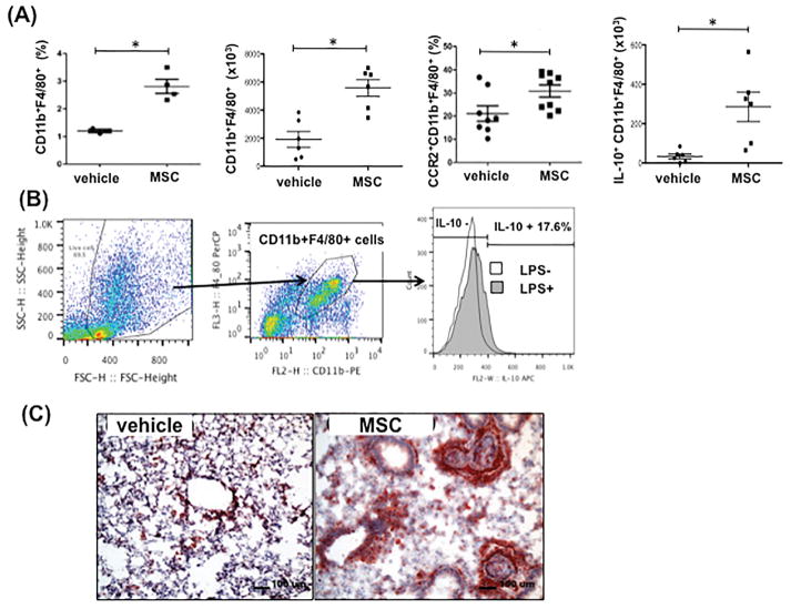
Figure 3. Monocyte and/or Macrophage Responses Following MSC Administration in the Lungs of Allergen Sensitized and Challenged Mice.
C57Bl/6 mice were sensitized and challenged with OVA, then injected i.v. with 2x10⁵ MSC. (A) Seven hours after MSC injection, lung cells from MSC-injected (MSC) or PBS-injected (vehicle) mice were isolated, then immunostained for identification of CD11b+F4/80+ (per total lung mononuclear cells), CCR2+CD11b+F4/80+ (per total CD11b+F4/80+ cells), or IL-10+CD11b+F4/80+ monocytes and macrophages (per total CD11b+F4/80+ cells) by flow cytometry (n=6 per group) (*p<0.05). (B) Gating strategy to detect IL-10-positive monocyte/macrophage in the lung tissue and a representative flow cytometry data. IL-10- levels in CD11b+F4/80 cells were compared following with or without LPS stimulation as describe in Materials and Methods. In (C), lung tissues from PBS-injected (left panel) and MSC-injected (right panel) mice were immunostained for identification of CD68+ monocytes and macrophages. Lungs from MSC-injected mice had prominent accumulations of CD68+ cells (red stain) around airways, compared to PBS-injected mice. Similar results were observed in 2 additional control and treated mice, processed similarly.

