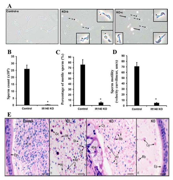
Figure 3. Disruption of Ift140 in male germ cells results in abnormal sperm morphology, reduced sperm number and mobility.
A. Morphological examination of cauda epididymal sperm collected from 3~4-month-old control (a) and conditional Ift140 KO mice (b–c) by light microscopy. Notice that compared to the control, much cell debris and very few sperms were observed in the conditional Ift140 KO mice under the same dilution. Sperms from the Ift140-deficient mice had bent (black arrows in c) or short tails (dashed arrows in b and c). Some sperms had swollen tail tip (asterisk in b and c). Vacuoles were present at the base of stunted flagella (arrowheads in b and c). B. Sperm count is significantly reduced in the KO mice. C. Comparison of percentage of motile sperm between the control and Ift140 KO mice. D. Analysis of sperm motility by calculating curvilinear velocity (VCL). Sperm VCL was largely decreased in the Ift140-deficient mice compared to the controls (* p < 0.05). E. Histology of the cauda epididymis from control (heterozygote) and conditional Ift140 mutant mice (KO). (a) Control epididymis showing highly concentrated epididymal sperm with aligned sperm tails (T) and normal sperm heads (H). Ep, epithelium. (b) KO epididymal lumen showing abnormal sperm heads (Ab), cytoplasmic bodies with and without sperm heads (H), short tails (St), residual bodies (Rb), kinked tails (Kt) and sloughed germ cells (Gc). (c) KO epididymis showing an area of abnormal epithelium (Abe). The lumen contains abundance of large cytoplasmic bodies (Cy), residual bodies (Rb) and numerous abnormally shaped sperm heads (Ab). (d) KO epididymis with normal epithelium (Ep), but a lumen containing scattered cytoplasmic bodies (Cy) and residual bodies (Rb), with no sperm heads present. Bars = 20 μm.

