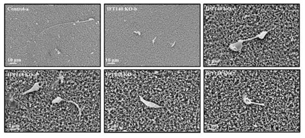
Figure 4. Examination of epididymal sperm by SEM.
(a) Representative image of epididymis sperm with normal morphology from a control mouse shows the sperm with a long, smooth tail and condensed head. (b–f) Representative images of epididymal sperm from a conditional Ift140-deficient mouse. A variety of morphologic abnormalities of sperm were observed. The sperm displayed amorphous heads (d, e), short/swollen flagella (b to f), uneven thickness of flagella (d), and other distorted shapes.

