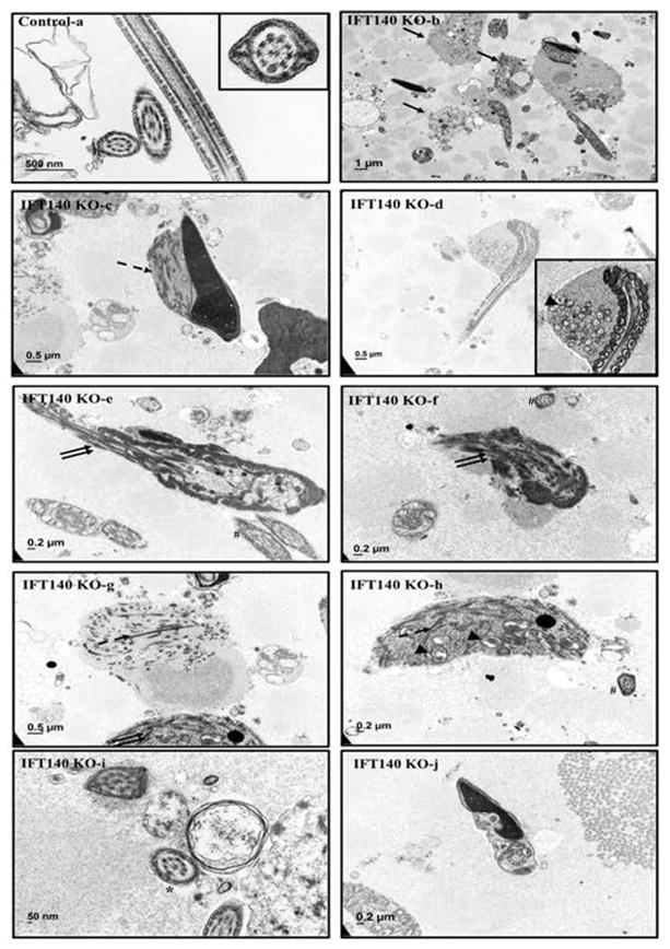
Figure 6. Analysis of epididymal sperm of the control and Ift140 KO mice by TEM.
TEM images of epididymal sperm from 4-month-old control and KO mice. Sperm from the control mice show normal ultrastructure (a). Few sperm were discovered in the collection from the KO mice. Instead, significant amounts of residual bodies (arrows in b) were present. The residual bodies and the very few sperm with redundant cytoplasmic components contained components for ODF and fibrous sheath (dashed arrow in c, g, h) and mitochondria (arrow head in d, h). Disorganized microtubule array (double arrows in e, f, g) were frequently seen. Some sperm lost “9+2” core axoneme structure (* in i); some have disorganized chromatin structure (j). Some sperm with apparently normal ultrastructure in cross-sections were still observed in the conditional Ift140 KO mice (# in e, f, h).

