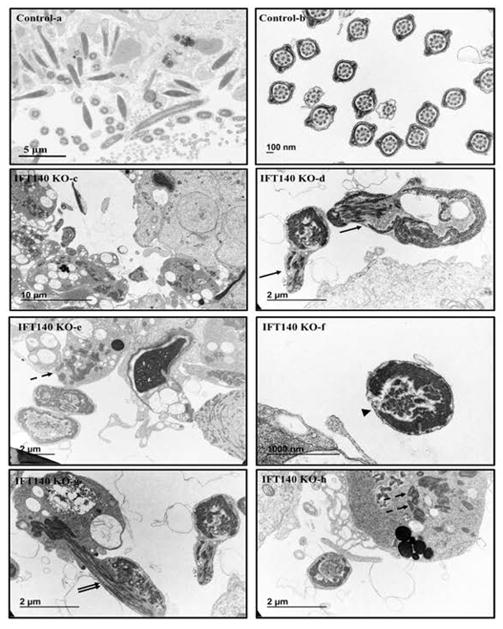
Figure 7. Analysis of testicular sperm of the control and Ift140 KO mice by TEM.
TEM images of testicular sperm from 4-month-old control and KO mice. In the control mice, sperm were easily discovered in the lumen of seminiferous tubules (a), and the ultrastructure was normal (b). However, few sperm were discovered in the KO (c), and the sperm usually have disorganized axoneme structure (arrows in d). The dashed-arrows in “e” and “h” point to the mitochondria materials in the residual bodies; the arrowhead in “f” points to an axoneme with disrupted “9+2” array; the double-arrows in “g” points to an axoneme with disorganized microtubules.

