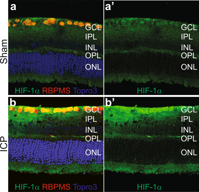
Figure 4.
ICP elevation causes increased HIF-1α expression. Representative retinal cross section images from Sham (a and a’) and ICP (b and b’) eyes stained for HIF-1α (green), RBPMS (RGCs, red), and Topro3 (nuclei, blue). HIF-1α expression is preferentially increased in the GCL after 2 weeks of ICP elevation (compare b’ to a’; see Table 3). GCL = retinal ganglion cell layer; IPL = inner plexiform layer; INL = inner nuclear layer; OPL = outer plexiform layer; ONL = outer nuclear layer.

