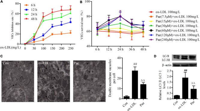
FIGURE 1.

Paeonol attenuates function of autophagy in ox-LDL-stimulated VECs. (A) The VECs were treated with serially diluted ox-LDL (50, 100, 150, 200, 250 mg/L) for different periods (6, 12, 24, 48 h) and subsequently the inhibitory rate as evaluate with the MTT assay. And it has a time- and dose-response relationship suppressed the growth of VECs. (B) VECs viability was detected by MTT assay after pretreatment with increasing concentrations of paeonol (7.5, 15, 30, 60, 120, 240 μM) for different period (6, 12, 24, 48 h), then treated with 100 mg/L ox-LDL for another 24 h. (C) Rat VECs autophagy by Electron Microscope (× 15000), arrows indicate autophagic vacuoles. Each sample counts six cells and showing the average number of double membrane vacuoles per cell. The data were expressed as mean ± SEM, n = 6. (D) Western blotting showing the expression of LC3 protein in VECs. Data were expressed as mean ± SEM, n = 3. ANOVA testing was performed; ##P < 0.01 vs. control; ΔΔP < 0.01 vs. ox-LDL group.
