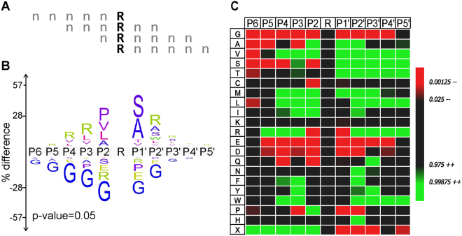
Figure 3.
Thrombin Substrate motif. (A) Schematic overview for alignment of peptides assuming Arg as P1. For peptides containing multiple Arg residues, the center-most Arg was aligned, with preference given to position 3 Arg residues in peptides containing Arg residues at position 3 and 4. (B) The iceLogo plot representing the relative frequency of every amino acid in the cleaved (top) and uncleaved (bottom) peptide pools following thrombin selection. (C) The iceLogo heatmap shows the preference for each amino acid at every position in the cleaved (green) or uncleaved (red) peptide pools. Residues at positions that do not appreciably populate either pool are also indicated (black).

