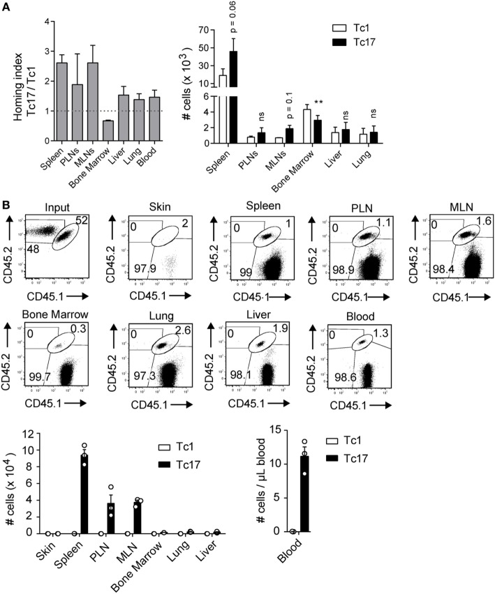
Figure 2.
Tc17 cells recirculate within secondary lymph nodes. (A) Tc1 and Tc17 lymphocytes were generated in vitro from CD45.2+ mice and stained with eFluor 680 (5 µM) and CMTMR (10 µM) respectively. Then the cells were co-transferred (0.5 × 10⁶ cells of each type) in CD45.1+ mice. Cell migration was evaluated 24 h later in several lymphoid and effector tissues. Homing index in each organ was calculated as (Tc17organ/Tc1organ)/(Tc17input/Tc1input), n = 3. (B) Tc1 cells (CD45.2+) and Tc17 cells (CD45.1+/CD45.2+) were generated in vitro and injected intradermally into congenic mice (CD45.1+). 32 days later, the number of transferred cells was analyzed in skin, spleen, peripheral lymph nodes (PLN), mesenteric lymph node (MLN), bone marrow, lung, liver, and blood of recipient mice, n = 3.

