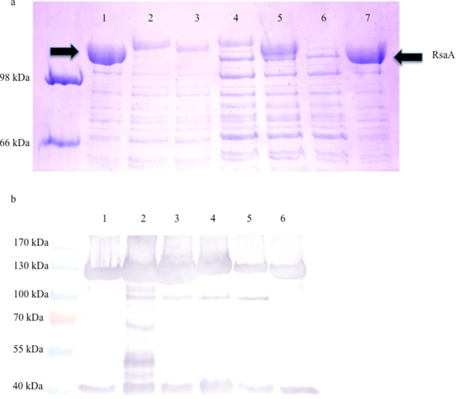
Figure 1.

Recombinant S-layer protein gel. Representative Coomassie blue stained 7.5% SDS-PAGE of normalized low pH extracted RsaA protein from C. crescentus strain JS 4038 containing RsaA plasmids. Arrows indicate the location of the S-layer protein (100 kDa), with size shifts visible to indicate expression of recombinant protein. 1) Cc-Control (no insert); 2) Cc-A1AT; 3) Cc-BmKn2; 4) Cc-Elafin; 5) Cc-Indo; 6) Cc-Indo2; 7) Cc-Control. The image has been brightened and cropped to minimize background. The original image has been provided to Scientific Reports and is available from the authors upon request. (b) Representative Western blot. RsaA was extracted from C. crescentus using a low pH method, normalized, run on 7.5% SDS-PAGE using a prestained protein ladder, and RsaA was detected by western blot using an anti-RsaA antibody. 1) Cc-Control (no insert); 2) Cc-A1AT; 3) Cc-BmKn2; 4) Cc-Elafin; 5) Cc-Indo; 6) Cc-Control.
